Steps in Wound Assessment
Accurate wound assessment is crucial for effective management and healing, particularly in burns and diabetic foot ulcers. Start by evaluating the wound size, depth, and location. Examine the tissue for signs of necrosis, infection, or granulation. Assess exudate type and volume, noting any odor. Measure the wound edges and check for undermining or tunneling. Use standardised tools like the Bates-Jensen Wound Assessment Tool (BWAT) for consistency. Document pain levels and patient symptoms. Regularly reassess wounds to monitor healing progress and adjust treatment plans accordingly. This systematic approach ensures comprehensive care and promotes optimal healing outcomes.
Initial Patient Evaluation
Initial patient evaluation is a critical first step in assessing wounds, especially for burns and diabetic foot care management. Start by gathering a comprehensive patient history. Ask about the wound’s onset, cause, and previous treatments. Investigate the patient’s medical history, focusing on conditions that could affect healing, such as diabetes, peripheral vascular disease, or immune deficiencies.
Next, perform a thorough physical examination. Assess the wound size, depth, and location. Identify the type of tissue present, noting any necrotic, granulating, or epithelialising tissue. Look for signs of infection, including erythema, warmth, swelling, and purulent discharge.
Steps in Initial Patient Evaluation:
- History Collection: Document the wound’s origin, duration, and any prior interventions.
- Medical History: Identify underlying conditions like diabetes, vascular diseases, and nutritional deficiencies that may impair healing.
- Physical Examination: Evaluate wound characteristics, including size, depth, and tissue type.
- Infection Signs: Observe for indicators of infection, such as redness, heat, and exudate.
Additionally, consider the patient’s nutritional status, as deficiencies can impede healing. Assess pain levels and psychosocial factors that may affect wound management compliance. Use standardised tools like the Bates-Jensen Wound Assessment Tool (BWAT) for consistent documentation.
More about Deepview
Learn more about our DeepView® technology
Request a Demo
Looking to learn more about DeepView® technology, or eager to see it in action?
Visual Inspection of the Wound
Visual inspection of the wound is a crucial step in wound assessment. Begin by assessing wound size and depth. Measure the length, width, and depth using a ruler or wound measurement tool. Document these dimensions accurately to track healing progress over time.
Next, evaluate the wound edges. Determine if they are attached or detached from the wound bed, rolled under (epibole), or macerated. Attached edges typically indicate healthy granulation tissue, while detached or rolled edges may suggest underlying issues with healing.
Examine the periwound skin thoroughly. Look for signs of maceration, which appears as white, waterlogged tissue, often caused by excessive exudate. Check for erythema, indicating possible infection or inflammation. Assess for induration, which can signal underlying infection or tissue damage. Evaluate for callus formation, particularly in diabetic foot ulcers, as this can impede healing and increase the risk of further tissue injury.
Consider the color, consistency, and odor of wound exudate. Clear or pale yellow exudate is usually normal, while purulent or foul-smelling discharge suggests infection. Regularly reassess these parameters to monitor changes and adjust the treatment plan accordingly.
By systematically evaluating wound size, depth, edges, and periwound skin, clinicians can develop a comprehensive wound management strategy. This approach ensures accurate monitoring of healing progress, early identification of complications, and timely adjustments to treatment protocols, ultimately promoting optimal wound healing outcomes.
Wound Bed Assessment
Begin by checking the wound bed for necrotic tissue, slough, and granulation. Necrotic tissue, often black or brown, needs removal to prevent infection and promote healing. Slough, which appears yellow or white, also hinders healing and requires debridement.
Next, evaluate the presence of granulation tissue. Healthy granulation tissue is red or pink and indicates good blood flow and healing. However, over-granulation, which can be excessive, may need intervention to ensure proper wound closure.
Identifying signs of infection or inflammation is critical. Look for erythema, increased warmth, and swelling around the wound, which may indicate inflammation or infection. Check for purulent exudate, characterised by a yellow, green, or brown color and often accompanied by a foul odor. Document these findings meticulously.
Assess pain and tenderness, as increased pain can signal infection or complications. Monitor systemic signs such as fever, which may require prompt medical intervention.
Additionally, observe for signs of biofilm, a slimy layer that can protect bacteria and impede healing. Biofilm may necessitate specialised treatments.
By thoroughly assessing the wound bed for necrotic tissue, slough, granulation, and signs of infection or inflammation, clinicians can develop an effective wound management plan. This approach promotes optimal healing, prevents complications, and ensures comprehensive care for patients with burns and diabetic foot ulcers.
Assessing Wounds Exudate
Assessing wound exudate begins by identifying the types of exudate, as they provide valuable insights into the wound’s condition:
- Serous exudate: Clear or pale yellow, generally indicates normal healing.
- Sanguineous exudate: Contains blood and may appear pink or red, suggesting new tissue formation or minor bleeding.
- Serosanguineous exudate: A mix of serous fluid and blood, common in healthy wounds.
- Purulent exudate: Thick, yellow, green, or brown, often has a foul odor, indicating infection.
Monitoring the volume and consistency of exudate is equally important. Excessive exudate can lead to maceration of the surrounding tissue, delaying healing. Insufficient exudate might indicate dehydration or poor circulation, impeding tissue repair. Use wound dressings that manage moisture effectively to maintain an optimal environment for healing.
Document changes in exudate characteristics regularly. Increased volume or a sudden change in color and consistency can signal infection or complications, necessitating a change in treatment strategy.
By carefully assessing the type, volume, and consistency of wound exudate, clinicians can make informed decisions about wound management. This practice promotes effective healing, prevents complications, and ensures comprehensive burn diagnosis.
More about Deepview
Learn more about our DeepView® technology
Request a Demo
Looking to learn more about DeepView® technology, or eager to see it in action?
Advanced Wound Assessment Techniques
Using a Wounds Assessment Checklist
A wound assessment checklist offers significant benefits for clinicians treating burns and diabetic foot ulcers. A standardised checklist ensures a systematic and thorough evaluation process, reducing the risk of overlooking critical details.
Key components of an effective checklist include:
- Wound Size and Depth: Measure accurately to track healing progress.
- Tissue Type: Identify necrotic, slough, and granulation tissues.
- Exudate: Assess type, volume, and consistency.
- Wound Edges and Periwound Skin: Check for signs of maceration or infection.
- Dressing Suitability: Evaluate if the current dressing supports optimal healing.
Implementing wound assessment guidelines enhances consistency, improves documentation, and ensures comprehensive wound management.
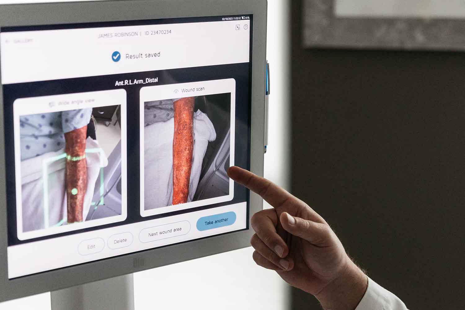
Utilising Technology in Wound Assessment
Digital tools and apps streamline the assessment process, providing accurate measurements and real-time documentation. These tools facilitate the tracking of wound size, depth, and tissue types, improving the consistency of evaluations.
Imaging techniques, such as thermography and laser Doppler, offer detailed visualisation of the wound bed and surrounding tissue. These methods identify areas of poor perfusion and infection early, allowing for timely intervention. Incorporating technology into wound management optimises dressing choices, enhances patient outcomes, and streamlines the overall treatment process.
Regular Monitoring and Documentation for Wound Management
Consistent documentation ensures accurate tracking of changes in wound healing over time, providing a comprehensive view of the wound’s progress.
Record key details, such as wound size, depth, tissue type, and dressing changes, during each assessment. By systematically documenting these parameters, clinicians can identify trends, detect complications early, and adjust treatment plans accordingly.
Factors Influencing Wound Healing
Patient-Specific Factors
Patient-specific factors significantly influence wound healing. Nutrition plays a crucial role; adequate protein, vitamins, and minerals are essential for tissue repair and immune function. Encourage patients to maintain a balanced diet to support the healing process.
Mobility impacts circulation and pressure relief. Promote regular, appropriate movement to enhance blood flow and reduce the risk of pressure ulcers. Comorbidities, such as diabetes, vascular diseases, and immune deficiencies, can delay healing. Manage these conditions effectively to improve outcomes.
Patient compliance is vital. Adherence to prescribed wound care routines, including dressing changes and medication, directly affects healing. Educate patients on the importance of following their care plan and provide support to address any barriers.
Wound-Specific Factors
Wound-specific factors significantly influence healing in patients. The location and type of wound are crucial. Wounds on areas with high mobility, such as joints, heal slower due to constant movement disrupting the healing process. Additionally, wounds on pressure points or areas with poor blood supply, like the feet in diabetic patients, are more prone to complications and slower healing.
The type of wound also affects healing. For instance, burns with necrotic tissue require more extensive debridement, delaying healing. In contrast, granulating wounds with healthy tissue heal faster with appropriate care.
Wound duration plays a critical role in healing potential. Chronic wounds, those present for more than four weeks, often develop biofilms and are more resistant to treatment. Early and aggressive intervention is essential to prevent wounds from becoming chronic.
By understanding and addressing these wound-specific factors during assessment and management, clinicians can optimise the dressing and treatment process, promoting effective healing and reducing the risk of complications.
Related Posts